Critical Things to Know Before Scheduling an Abortion
At Heartbeats, we know it’s your life that will be impacted by the decision you make about your pregnancy. So, before you pay someone to perform an abortion, it is your right to know all of your options and have all of the information you need to make an educated, and safe, decision. While for-profit clinics and hospitals are often driven more by money than concern for the patient, we exist solely because we care about you, without making a profit.
Our knowledgeable, compassionate staff are committed to thoroughly and honestly sharing the information you need to make an informed decision, including answering these three critical questions before scheduling an abortion.

Is Your Pregnancy Viable?
A viable pregnancy means you are carrying a baby that has a reasonable chance to develop fully and survive outside the womb. A non-viable pregnancy, then, means the fetus has either died or has no chance of being born alive and living outside the womb1 . Some non-viable pregnancies, such as an ectopic pregnancy (a pregnancy that is growing outside of the uterus), can pose a significant risk to the mother and cannot be addressed through abortion. For this reason, having an ultrasound prior to scheduling an abortion is critical, as it is the only way to definitively determine viability. At Heartbeats, we can perform this ultrasound free of charge.

Learn More About Non-Viable Pregnancies
Non-Viable Pregnancies
Again, a non-viable pregnancy means that the baby has zero chance of surviving outside the womb. While there are strict medical guidelines for determining pregnancy viability, it is important that you are fully informed before moving forward with any medical procedure.
You’re much more likely to have a failed, or non-viable, pregnancy in the first trimester (the first 0-13 weeks of pregnancy)2. Any suspicion of a non-viable pregnancy should be discussed with your medical professional and all options explored before action of any kind is undertaken. A second opinion is always a good idea. While 10-20% of known pregnancies end in miscarriage3, there are other pregnancies that continue despite being non-viable and can potentially cause health risks. With that in mind, here are some of the most common causes of non-viability that may be detected through an ultrasound performed at 6 weeks gestation or later.
- No heartbeat. Keep in mind that if the gestational age of the pregnancy has not definitively been determined, it may be too early to detect a fetal heartbeat. Waiting a week or two and repeating the vaginal ultrasound may be in order. If a second ultrasound does not show a heartbeat, it could mean that you have miscarried or that the baby has died in utero. There could be a variety of reasons that the baby failed to thrive and develop. Consult with your medical professional regarding the need for a procedure known as dilation and curettage (D&C) or other method to ensure the safe and full expulsion of the fetus, placenta and pregnancy tissue from the uterus. During a D&C, the cervix is dilated and the contents of the uterus are removed using suction and/or a looped tool called a curette4.
- Ectopic pregnancy. This condition occurs when the fertilized egg implants outside of the uterus, most often in the fallopian tubes. An ectopic pregnancy affects 1% to 2% of all pregnancies and poses a significant threat to women of reproductive age. If left undiagnosed or untreated, the fetus can grow until it ruptures the fallopian tube, which will cause heavy internal bleeding in the abdomen and may lead to shock. It is the leading cause of maternal death during the first trimester of pregnancy and is responsible for 9% of pregnancy-related deaths in the United States5.
To prevent these life-threatening complications, the ectopic tissue must be removed using medication, laparoscopic or abdominal surgery. The method depends on your symptoms and when the ectopic pregnancy is discovered6.
- Anembryonic Gestation/Blighted Ovum. When a fertilized egg attaches to the uterine wall, it begins to develop a gestational sac around itself. In the case of anembryonic gestation, or blighted ovum, the gestational sac continues to grow, but the egg inside it does not, and it never develops into an embryo. This condition is believed to be the result of chromosomal abnormalities and often ends in miscarriage before or shortly after the woman becomes aware she is pregnant7.
If a miscarriage does not occur, the condition can be detected during an ultrasound that shows the gestational sac to be empty. At that point, your doctor may recommend waiting for a natural miscarriage to occur or suggest a D&C.
- Molar Pregnancy. This is a rare complication (1 in 1,000 pregnancies) that can present as either a complete or partial molar pregnancy. In a complete molar pregnancy, the placental tissue develops abnormally, becoming swollen and forming fluid-filled cysts that may appear like grapes on an ultrasound. A fetus does not form in this type of molar pregnancy because the egg that is fertilized is empty, meaning that the genetic material comes solely from the father’s sperm. A partial molar pregnancy, on the other hand, may contain both normal and abnormal placental tissue that forms simultaneously. A fetus may also form, but it is rarely able to survive because the abnormal tissue overtakes the fetus and/or because two sperm fertilize the same egg, thus providing two sets of male chromosomes, or two sets of the father's genetic material. If a doctor suspects a molar pregnancy, blood tests and an ultrasound will usually be ordered. If the pregnancy doesn’t end in miscarriage, other treatment options will be explored8.
In extremely rare instances, an embryo does develop and survive into the late weeks of a molar pregnancy, so while considered a non-viable pregnancy, it is always important to get conclusive evidence before moving forward. Women who are younger than 20 or older than 35 are at slightly higher risk of having molar pregnancies. There is also a chance that the molar pregnancy can develop into a cancerous tumor and spread beyond the uterus if not treated successfully9,10.
How Far Along Are You?
The gestational age of the fetus, or number of weeks since conception, is a key factor in determining the type of abortion you will receive, as well as its cost. Even though many women have a general idea of the date of their last period, the exact time the pregnancy began is an estimation. An ultrasound is the only way to definitively identify the true age and size of the fetus. In fact, without it, you could be offered the wrong type of abortion. A chemical abortion (the abortion pill), for example, could be recommended when you are actually past the 10-week window for that procedure’s safety or effectiveness. For this reason, a tele-medicine consultation is insufficient, as it cannot provide proof of pregnancy, proof of gestational age, or proof of a viable pregnancy, potentially putting you at risk. At Heartbeats, we personally provide all of this information at no cost to you.

Learn More About Types of Abortions
Do You Have an STD?
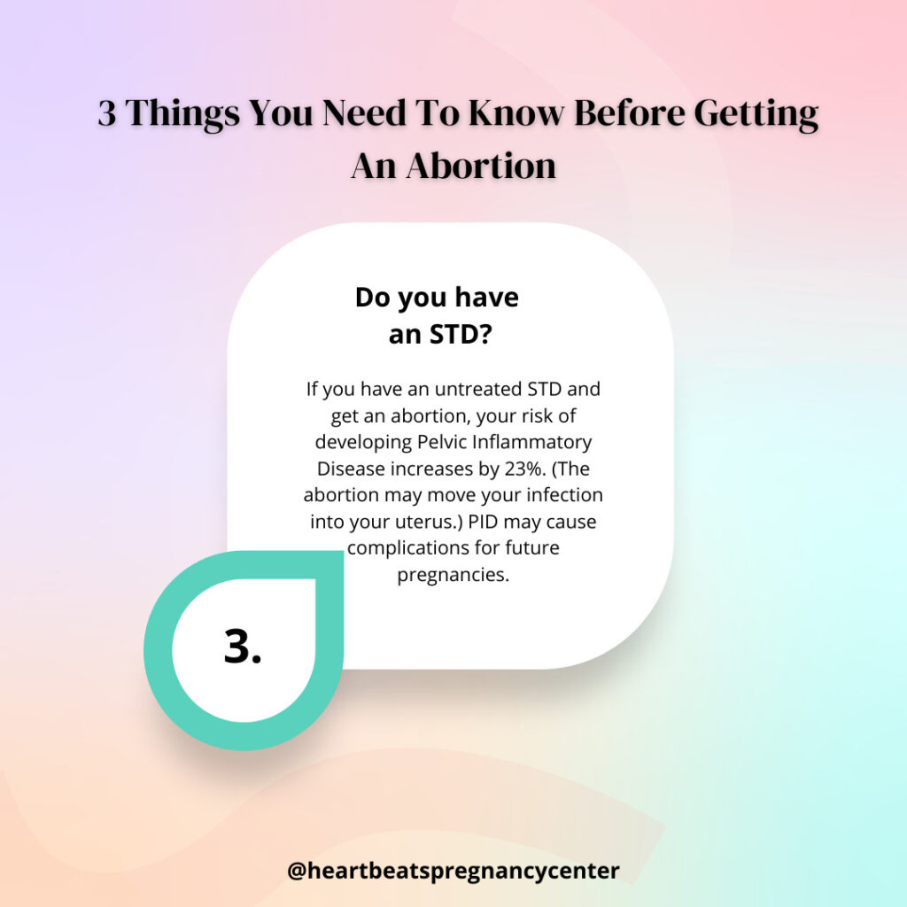
You may wonder what having an STD has to do with getting an abortion, but it is extremely important. If you have an STD, especially one of the two most common, chlamydia or gonorrhea, and aren’t treated before having an abortion, your risk of developing Pelvic Inflammatory Disease (PID) increases by 23% if the cervical infection is forced up into the uterus during the medical procedure25. PID increases your chances of having a future ectopic pregnancy, can decrease fertility, and can cause life-long pelvic inflammation and pain26. Testing is especially important because these STDs can be present without any symptoms. Other STDs, such as cervical syphilis27, HIV/AIDS28, and Human PapillomaVirus (HPV)29, also need to be tested for early in pregnancy, regardless of your pregnancy intentions, as they can pose significant risks to your health.
The majority of abortion facilities do not test for STDs prior to performing an abortion procedure. If they do, they charge an additional fee. At Heartbeats, we can confidentially have you tested and treated for these STDs at no charge. Results of STD testing are usually available within one week.

Learn More About STDs that Impact Abortion
Get Your Pre-abortion Screening
At Heartbeats, we are here to give you the answers to these three critical questions before undergoing an abortion. Our no-cost pre-abortion screenings include a pregnancy test, an ultrasound and STD testing all performed by a licensed medical professional. Call us at 740-349-7558 in Newark or at 740-450-5437 in Zanesville to schedule your screening today.

References
- Clement EG, Horvath S, Mcallister A, Koelper NC, Sammel MD, Schreiber CA. The language of first-trimester nonviable pregnancy: Patient-reported preferences and clarity. Obstet Gynecol.2019;133(1):149-154. doi:10.1097/AOG.0000000000002997
- (2021, June 15). A Determined Look into Non-Viable Pregnancy: Heartbreak and The Way Forward | Mommy Labor Nurse. Mommy Labor Nurse | Educating Expecting Parents About What’s To Come! https://mommylabornurse.com/non-viable-pregnancy/
- Miscarriage – Symptoms and causes. (2019, July 16). Mayo Clinic. https://www.mayoclinic.org/diseases-conditions/pregnancy-loss-miscarriage/symptoms-causes/syc-20354298
- How a D&E Differs From a D&C. (2020, November 8). Verywell Family. https://www.verywellfamily.com/what-is-dilation-and-evacuation-d-e-for-miscarriage-2371460
- Hillson, B. B. J. H. M. (2014, July 1). Diagnosis and Management of Ectopic Pregnancy. American Family Physician. https://www.aafp.org/afp/2014/0701/p34.html
- Ectopic pregnancy – Diagnosis and treatment – Mayo Clinic. (2020, December 18). Mayo Clinic. https://www.mayoclinic.org/diseases-conditions/ectopic-pregnancy/diagnosis-treatment/drc-20372093
- Blighted Ovum: A Non-Viable Pregnancy With No Obvious Symptoms. (2020, March 25). Verywell Family. https://www.verywellfamily.com/understanding-blighted-ovum-2371492
- How Are the Symptoms of a Molar Pregnancy Treated? (2020, October 25). Verywell Family. https://www.verywellfamily.com/molar-pregnancy-causes-symptoms-and-treatment-2371405
- Symptoms & Treatment For Molar Pregnancy Cancer. (2020). Www.Pregnancy-Baby-Care.Com. http://www.pregnancy-baby-care.com/molar-pregnancy/molar-pregnancy-cancer.html
- Feature Editor. (2019, August 28). Molar Pregnancy – What is it and Why Does it Happen?Com. https://pregged.com/molar-pregnancy/
- Abortion Pills – First Trimester Medical Abortion. (Accessed October 2021). abortionprocedures.com. https://www.abortionprocedures.com/abortion-pill/#1465365763472-92a2fc8d-9104.
- Center for Drug Evaluation and Research. (2021, April 13). Questions and Answers on Mifeprex. U.S. Food and Drug Administration. https://www.fda.gov/drugs/postmarket-drug-safety-information-patients-and-providers/questions-and-answers-mifeprex
- Incidence of Emergency Department Visits and Complications. . . : Obstetrics & Gynecology. (2015). LWW. https://journals.lww.com/greenjournal/Fulltext/2015/01000/Incidence_of_Emergency_Department_Visits_and.29.aspx
- Niinimäki, M. (2009). Immediate complications after medical compared with surgical termination of pregnancy. PubMed. https://pubmed.ncbi.nlm.nih.gov/19888037/
- Elective abortion: Does it affect subsequent pregnancies? (2020, September 19). Mayo Clinic. https://www.mayoclinic.org/healthy-lifestyle/getting-pregnant/expert-answers/abortion/faq-20058551?reDate=15102021
- Dilation and Curettage (D&C): Treatment, Risks, Recovery. (2021, March). Cleveland Clinic. https://my.clevelandclinic.org/health/treatments/4110-dilation-and-curettage-d–c
- Lohr, Patricia A. “Surgical Abortion in Second Trimester”, Reproductive Health Matters, May 2008, 156. ncbi.nlm.nih.gov/pubmed/18772096.
- D&E Abortion – Second Trimester. (Accessed October 2021). abortionprocedures.com. https://www.abortionprocedures.com/#1466802055946-992e6a14-9b1d.
- Bartlett, L. A. (2004, April). Risk factors for legal induced abortion-related mortality in the United States. PubMed. https://pubmed.ncbi.nlm.nih.gov/15051566/
- Fergusson, David M with Joseph M. Boden and L. John Harwood. “Does abortion reduce the mental health risks of unwanted or unintended pregnancy? A re-appraisal of the evidence.” Australian & New Zealand Journal of Psychiatry, Sept. 2013, Vol. 47, No. 9, pp. 819-827. http://www.ncbi.nlm.nih.gov/pubmed/23553240 .
- Darney, P.D., et al. “Digoxin to facilitate late second-trimester abortion: a randomized, masked, placebo-controlled trial.,” Obstetrics and Gynecology, Vol. 97, Issue 3, Mar.2001, pp. 471-476. ncbi.nlm.nih.gov/pubmed/11239659 .
- Induction Abortion – Third Trimester. (Accessed October 2021). abortionprocedures.com. https://www.abortionprocedures.com/induction/#1466802482689-777ef64c-4991.
- Dolle, J. M. (2009, April 1). Risk Factors for Triple-Negative Breast Cancer in Women Under the Age of 45 Years. Cancer Epidemiology, Biomarkers & Prevention. https://cebp.aacrjournals.org/content/18/4/1157.full
- Induction of fetal demise before abortion. (in press). Society of Family Planning. https://www.societyfp.org/_documents/resources/InductionofFetalDemise.pdf
- L, W., T, P., & J, S. (1982, September 1). Significance of cervical Chlamydia trachomatis infection in postabortal pelvic inflammatory disease. Abstract – Europe PMC. https://europepmc.org/article/med/7121913
- Pelvic Inflammatory Disease – CDC Fact Sheet. (1999). CDC. https://www.cdc.gov/std/pid/stdfact-pid.htm
- STD Facts – Syphilis. (2017, June). CDC. https://www.cdc.gov/std/syphilis/stdfact-syphilis.htm
- About HIV/AIDS | HIV Basics | HIV/AIDS | CDC. (2021, June). CDC. https://www.cdc.gov/hiv/basics/whatishiv.html
- STD Facts – Human papillomavirus (HPV). (2021, January). CDC. https://www.cdc.gov/std/hpv/stdfact-hpv.htm
- L, W., T, P., & J, S. (1982, September 1). Significance of cervical Chlamydia trachomatis infection in postabortal pelvic inflammatory disease. Abstract – Europe PMC. https://europepmc.org/article/med/7121913
- STD Facts – Chlamydia. (2014, January). CDC. https://www.cdc.gov/std/Chlamydia/stdfact-Chlamydia.htm
- STD Facts – Gonorrhea. (2014, January). CDC. https://www.cdc.gov/std/gonorrhea/stdfact-gonorrhea.htm